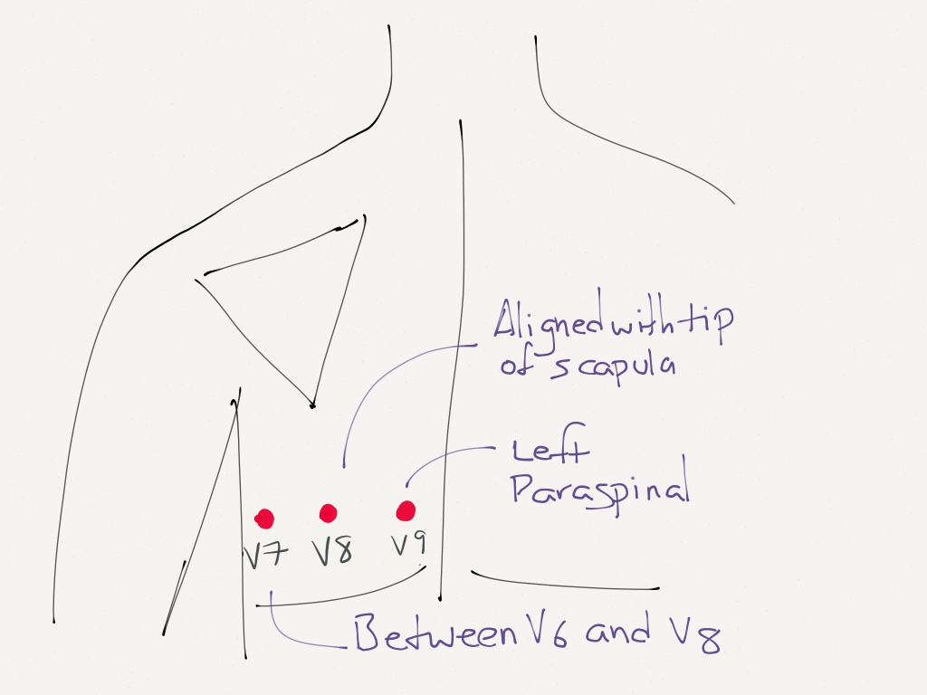
The Right Ventricle The Posterior aspect of the left ventricle. Placement of posterior leads V7-V9.

Diagnostics Alternative Ekg Leads Taming The Sru
V9 Left paraspinal region in the same horizontal plane as V6.

. From here the next step is appropriate lead placement for verification. I would like to thank Dr. Once the electrocardiogram with posterior leads has been made you must write the word Posteriors in the EKG header and overwrite leads V7 V8 V9 on the leads that have been replaced by.
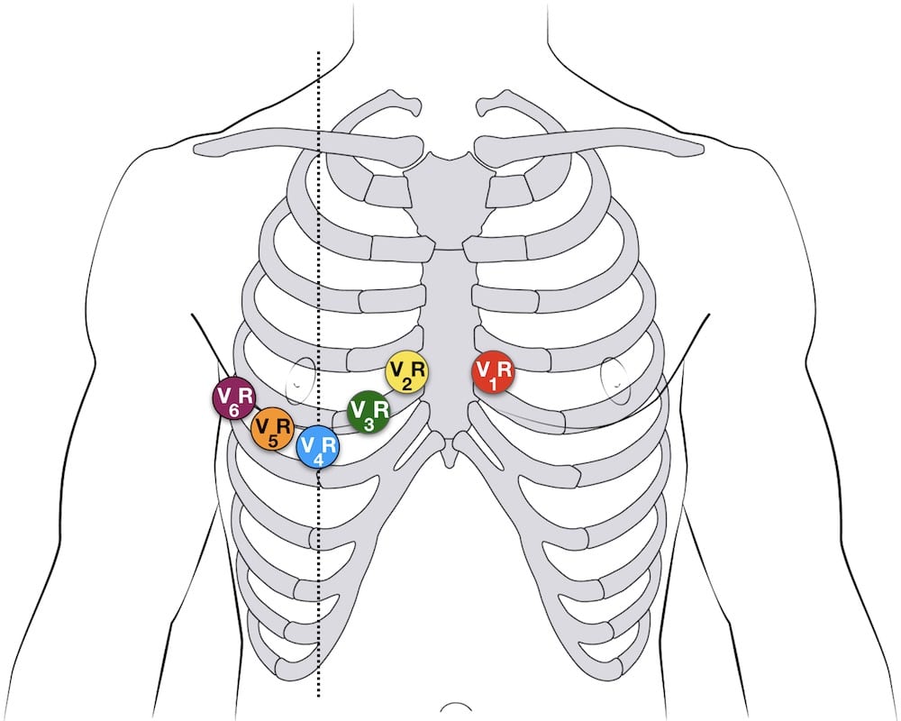
Therefore in patients presenting with acute chest pain and ST depression in the 12-lead ECG concomitant posterior ST elevation may be. At a minimum lead V4 should be placed on the 5th intercostal mid-clavicular exact opposite of the regular left side placement if an inferior infarct was originally seen in leads II III and AVF. Lead Placement for Posterior ECG.
The posterior electrodes V8 and V9 are placed on the patients backV8 at the tip of the left scapula and V9 in an intermedi- ate position between lead V8 and the left paraspinal muscles. What is a posterior lead in ECG. V7 Left posterior axillary line in the same horizontal plane as V6.
26 Votes V7 is placed at the posterior axillary line in the same horizontal plane as V6. Posterior Ventricular leads V7 V8 V9. This blog aims to disrupt how medical providers and trainees can gain public access to high-quality educational content while also engaging in a dialogue about best-practices in EM and medical education.
V8 Tip of the left scapula in the same horizontal plane as V6. V7 is located at the same horizontal line as V4R ie 5th ICS on the posterior axillary line use the V4 electrode. There are two areas of the heart for which none of the standard 12 leads monitor the electrical activity.
The leads V4-V6 are removed and substituted for V7-V9 as shown below. Reposition cable to prevent electrodes from pulling away from patient. V9 same horizontal line as V4R left paraspinal border use V6 electrode.
These areas are most accurately monitored by the placement of special leads V4R V7 V8 V9. V9 is placed in the left paraspinal region in the same horizontal plane. Lastly a right sided 12-lead ECG placement allows you to detect a right sided infarct.
The commonest additional leads are the posterior leads V7 V8 and V9 that view the posterior and lateral walls and the right V4 RV4 that examine the right ventricle. It is also helpful for future clinicians if you note in your read that it is a posterior ECG. Remeber your coronary artery anatomy.
Therefore the use of the 15-lead ECG may confirm the STEMI diagnosis while determining its actual extent. This can be remembered by V789 staying on the same plane as previous V6 with V8 placement to the tip of the scapula. Gemma Morabito MedEmIt for the idea of this post and Amal Mattu amalmattu for these.
V4V7 V5V8 and V6V9. At the same level as electrodes V6 the left paravertebral line. V7-V9 Ensure leads are properly connected.
V8 same horizontal line as V4R mid subscapular line use V5 electrode. Level with V7 at mid-scapular line V9. ST elevation in leads V7 V8 and V9 is uncommon in patients presenting with subendocardial ischaemia.
Leads I II and III. Level with V8 just left of vertebral line Special Lead Placement. When doing a right-sided EKG what is the placement of the leads.
What are the lead groups that represents Einthovens Triangle. Once the electrocardiogram with posterior leads has been made you must write the word Posteriors in the EKG header and overwrite leads V7 V8 V9 on the leads that have been replaced by posterior leads. V8 is placed at the tip of the left scapula in the same horizontal plane.
V9 is placed in the left paraspinal region in the same horizontal plane. Where should a CCMA place the electrodes for leads V7 V8 and V9. Medical health heart and cardiovascular diseases.
V8 Tip of the left scapula in the same horizontal plane as V6. Read full answer here. V7 is placed at the posterior axillary line in the same horizontal plane as V6.
All thats needed to diagnose a STEMI in V7V8V9 is. Left posterolateral chest leads V7 V8 V9 helped distinguish the multiple causes of tall R waves in V1 andor V2 diagnosed true posterior myocardial infarction when standard leads did not and identified the presence or absence of posterior injury in patients with inferior infarction. On most EKg machines the labels areno automatically changed so it is important to cross out the labels for V4-V6 and write in V7-V9.
V8 is placed at the tip of the left scapula in the same horizontal plane. Pick up V4 V5 V6 and replace with V7 V8 V9 V7. ELECTROCARDIOGRAM ALTERNATE LEAD PLACEMENTS RIGHT SIDED OR V7 V8 V9 2140712 Procedure Posterior V 7-9 ECG 1 Perform a routine 12 lead ECG with regular limb and chest lead placement.
Left posterior axillary line V8. Troubleshooting Artifact Prepare the patients skin and apply new. Divide 1500 by the number of small boxes between two R waves.
2 Reposition the chest electrodes per the attached diagram for V 7 V 8 V 9 on the patients back. Left paraspinal region Look for ST elevations in V7 V8 V9 on your p osterior EKG. To clarify leads will equal.
Leads V7-9 are placed on the posterior chest wall in the following positions see diagram below. 415 45 Views. Leads V7-9 are placed on the posterior chest wall in the following positions.
An additional electrode V7 may also be used and is placed on the posterior axillary line equidistant from electrode V8 Fig. Placement of Posterior Leads. V7 Left posterior axillary line in the same horizontal plane as V6.
Left tip of scapula V9. A cheap and easy way to diagnose a posterior MI is flipping the ECG over and looking at leads V1 V3 in the light but using posterior leads V7 V9 will more accurately diagnose patients with posterior MI. V9 Left paraspinal region in the same horizontal plane as V6.
When do you add additional leads such as V7 V8 V9 V4R. At the same level as electrodes V6 the left paravertebral line.

Posterior Ekg Lead Placement Emergency Nursing Nursing Notes Ekg

Ecg Lead Positioning Litfl Ecg Library Basics

Posterior Electrode Placement V7 Is Placed In The Left Posterior Download Scientific Diagram

Electrocardiographic Diagnosis Of Remote Posterior Wall Myocardial Infarction Using Unipolar Posterior Lead V9 Chest

How To Not Miss A Posterior Myocardial Infarction Em Daily

Brmbar Osnovatel Ponyakoga Ponyakoga V7 V8 V9 With 12 Chanel Ekg Teknologipembelajaran Com
Ecg Lead Positioning Litfl Ecg Library Basics
Lead Placement For Posterior Ecg Resus Review
0 komentar
Posting Komentar